HSIEH-WILSON LAB
  • Home
  • Research
    • Overview
    • Decoding HS
    • Neuroplasticity & CS
    • O-GlcNAc & PTMs
    • Glycan structure ID
  • People
  • Meet Linda
  • Publications
  • Photos
  • Contact

Glycan structure identification at the single-cell level

The brain is a highly complex organ, comprised of many different cell types and subclasses of cells. Our understanding of this cellular complexity has rapidly expanded with the advent of single-cell methods such as single-cell RNA sequencing (scRNA-seq). Single cell methods have been powerful in advancing our understanding of genetic molecular signatures, however, analogous methods to examine the glycan molecular signature of single cells have been lacking. Our lab has expanded our chemoenzymatic labeling strategies to specifically detect and profile the glycan signature of individual cells. In multiplexing with scRNA-seq, thus classifying cells by both gene expression and glycan expression profiles, we identify specific glycan structures, biosynthesis enzymes, glycoproteins, binding proteins, and cell types that contribute to brain processes such as learning, memory, and disease.
Picture
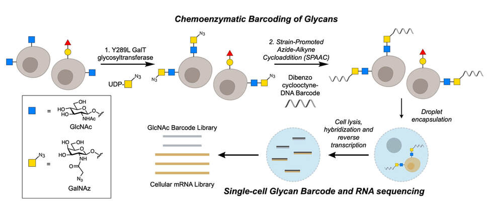
Overview of chemoenzymatic labeling of cell-surface glycans
Fucose a(1-2) galactose glycans
Fucose-α(1-2)-galactose (Fucα(1-2)Gal) is a glycan of interest in learning and memory formation, but the structural complexity of fucose-containing glycans has made their study difficult. Although there is an antibody against Fucα(1-2)Gal, it binds too weakly to enrich fucose-containing glycoproteins, further limiting efforts to study this glycan. The Hsieh-Wilson lab has developed a method for chemoenzymatic labeling of Fucα(1-2)Gal which, when combined with high-throughput mass spectrometry, led to the first proteomic analyses of fucosylated glycoproteins and binding partners from the mammalian brain. Our study suggested that fucose plays important regulatory roles in phenomena such as cell adhesion and synaptic plasticity, but the specific mechanisms involving fucose in these phenomena, and how they factor into learning and memory, remain unclear.
Chaubard et al., "Chemoenzymatic Probes for Detecting and Imaging Fucose-α(1-2)-Galactose Glycan Biomarkers," J. Am. Chem. Soc. 2012. DOI: https://doi.org/10.1021/ja211312u 
Picture
Detection of Fucα(1-2)Gal-containing glycans on neurons with an antibody.
SUGAR-seq
Leveraging our existing chemoenzymatic labeling techniques, our lab has developed a method to detect and profile the levels of specific glycans on individual cells. When profiled alongside gene expression, we identify glycan-mediated cellular processes at single-cell resolution and establish a framework for the study of cellular processes within cell types and subpopulations.
For example, in establishing cellular glycan signatures across samples, we identify changes in glycosylation among different cell cycle phases, cell types, and disease phenotypes.
Picture
Profiling glycans at the resolution of single cells.
Go back
Picture
Vertical Divider
Picture
Hsieh-Wilson Laboratories
1st Floor, Norman W. Church Laboratory for Chemical Biology / Caltech
Prof. Linda C. Hsieh-Wilson: [email protected]
  • Home
  • Research
    • Overview
    • Decoding HS
    • Neuroplasticity & CS
    • O-GlcNAc & PTMs
    • Glycan structure ID
  • People
  • Meet Linda
  • Publications
  • Photos
  • Contact